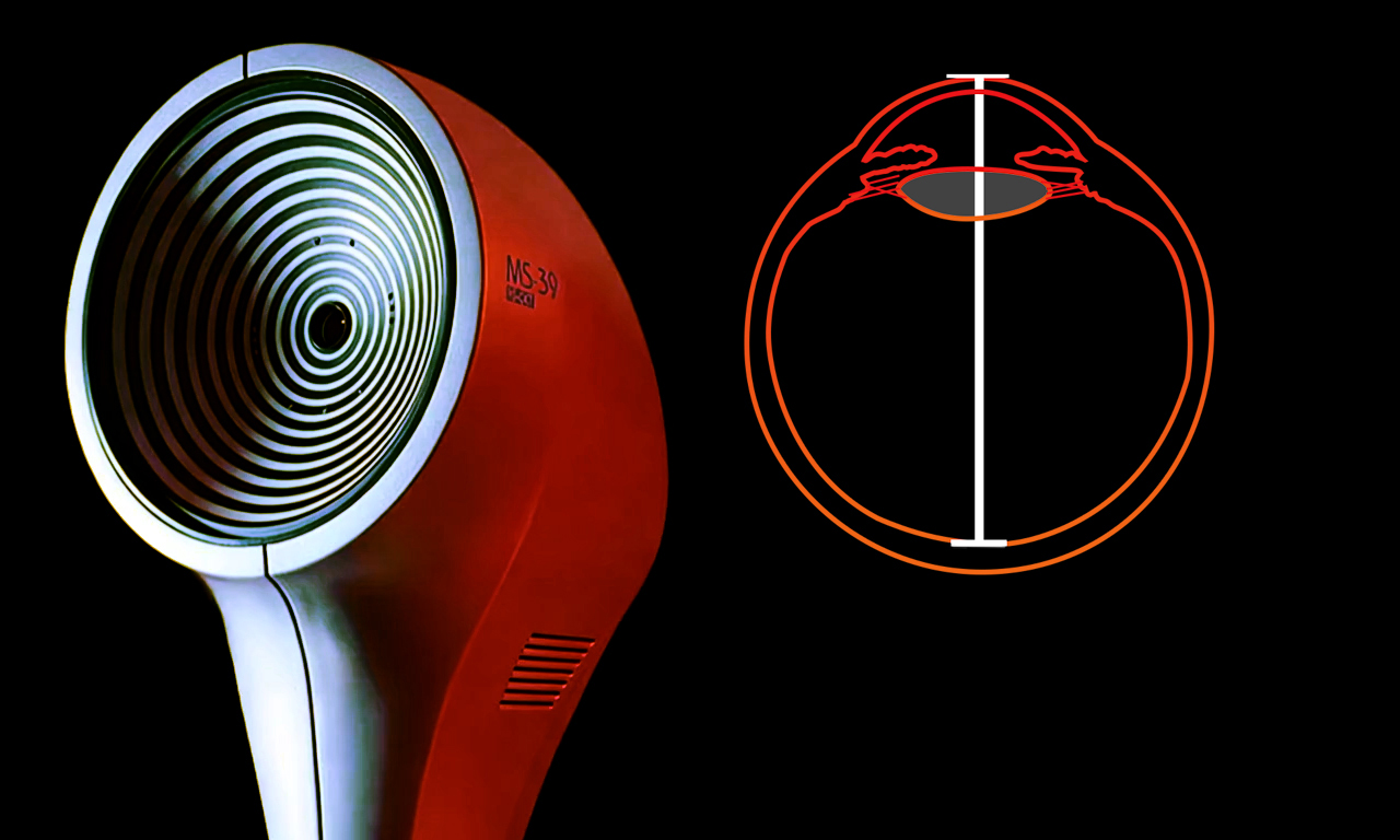

MS-39 AS-OCT
Is the most advanced device for the analysis of the anterior segment & anterior chamber of
the eye. MS-39 combines Placido disk corneal topography, with high resolution OCT-based anterior segment
tomography. The clarity of the cross-sectional images, with a 16 mm diameter, along with the many details of
the cornea structure and layers revealed by the MS-39, will be appreciated by anterior segment specialists.
MS-39 provides information about the epithelium layer, pachymetry, elevation, curvature and dioptric power of both corneal surfaces.
In addition to anterior segment clinical diagnostics, MS-39 can be used in corneal surgery for refractive
surgery planning. An IOL calculation module is also available, based on Ray-Tracing techniques, Additional
tools allow MS-39 to perform accurate pupil diameter measurements and the advanced analysis of tear film.
The CSO MS39 examines the smallest hidden details such as stromal haze, ICRS depth, deep demarcation line, historical epithelium details, remodelling information and detecting masked abnormalities.

SD-OCT Axial Length
Spectral Domain OCT high accuracy measurement

SD-OCT Axial Length
Spectal Domain OCT high accuracy measurement
HD ANTERIOR SEGMENT & CHAMBER
High-resolution section images on a diameter of 16 mm
CORNEAL SECTION
The sharpness of the high-resolution section images on a diameter of 16 mm, together with the many details of the structure and the cornea layers brought to light by the instrument, are the most extraordinary features and appreciated by the specialists of the anterior segment (corneal and epithelial). The device provides pachymetry, elevation, curvature and power information for both corneal surfaces down to an unequalled 3.5µ of precision.

EPITHELIAL AND STROMAL MAP

MS-39 includes the advanced measurement of the epithelial and stromal layer. The epithelial masking effect is known, so knowledge of its morphology is very useful assess abnormalities of the corneal surface. The information about the epithelium provided by MS-39 is crucial for the surgical planning in refractive surgery and for the management of keratoconus.
CORNEAL ABERROMETRY
Aberrometric analysis offers a complete overview of the corneal aberrations. It is possible to select the contribution of the anterior, posterior or total cornea for different pupil diameters. The OPD/WFE maps and the visual simulations (PSF, MTF, image convolution) can help the clinician in understanding or explaining the patient’s visual problems.

CRYSTALLINE LENS BIOMETRY

In order to more accurately determine the ELED, and consequently to refine the IOL (intra-ocular lens) calculation, MS-39 provides an acquisition mode to measure the crystalline lens thickness, its distance from the cornea and its equator.
ADVANCED KERATOCONOUS SUMMARY
Keratoconous screening provides the clinician with important information about the patient's cornea. Understanding this can help prevent complications associated with ectasia before corneal surgery is undertaken.

TECHNICAL DATA
- Data transfer USB 3.0
- Power supply external power source 24 VDC In: 100-240Vac - 50/60Hz - 2A - Out: 24Vdc - 100W
- Power cable IEC C14 plug
- Dimensions (HxWxD) 505 x 315 x 251mm
- Weight 10.4Kg
- Chin rest movement 70mm ± 1mm
- Minimum height of the chin cup from table 23cm
- Base movement (xyz) 105 x 110 x 30mm
- Working distance: 74mm
- LIGHT SOURCES
- Placido disk illumination Led @635nm
- OCT source SLed @845nm
- Pupillographic illumination Led @950nm
- TOPOGRAPHY
- Placido disk rings 22
- Measured points 31232 (anterior surface) 25600 (posterior surface)
- Topographic covering 10mm
- Dioptric measurement range from 1D to 100D
- Measurement accuracy Class A according to UNI EN ISO 19980-2012
- TOMOGRAPHIC SECTION
- Image field 16mm x 8mm
- Axial resolution >3.6µm (in tissue)
- Transversal resolution 35µm (in air)
- Image(s) resolution Keratoscopy (640x480) + 25 radial scans on a 16mm transversal field (1024 A-scan)
- Section: on 16mm (1600 A-scan) on 8mm (800 A-scan)
- MINIMUM SYSTEM REQUIREMENT
- PC: 4GB RAM - Video Card 1 GB RAM - resolution 1024 x 768 pixels - USB 3.0 type A Operating system: Windows XP, Windows 7 and Windows 10 (32/64 bit).